Central line placement helps provide essential care for hospitalized and critically ill patients. But like most medical procedures, it has a few complications that can be fatal, including hematoma, pneumothorax, infection, bleeding, DVT and extravasation.
Hence, physicians and other medical professionals must understand the proper techniques, particularly the catheter tip’s correct positioning, to reduce complications and enhance patient outcomes.
Fortunately, ultrasound guidance, new catheter designs, and standardized techniques have made the procedure safer and more commonplace in the ER or intensive care unit.
In this piece, we look at how techniques like Peres’ height formula and the topographical method can aid in determining the central line depth of insertion for different sites. But first, let’s understand what a central line placement is, why it’s essential, and how it’s done.
What Is a Central Line Placement and Why Is It Necessary?
A central line is a small catheter inserted in a vein for long-term intravenous drug therapy, central hemodynamic monitoring, or parenteral nutrition. Its placement is required when patients need drugs administered through their veins for long periods or when they need kidney dialysis through a dialysis catheter.
In these situations, inserting a central venous catheter makes the process easier and less painful than inserting needles in a patient’s veins every time they require therapy.
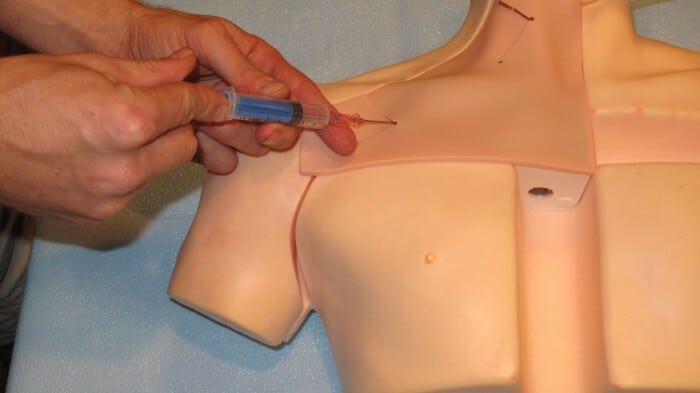
How Is a Central Line Placement Procedure Done?
Usually, physicians, physician assistants, nurse practitioners, or trained nurses perform central line placement procedures using ultrasound guidance. During insertion, the operator will insert a tiny catheter in the patient’s vein either in the internal jugular vein, the subclavian vein or the femoral vein and then secures it in place.
The catheter will remain intact for drug treatment or dialysis.
Determining Central Line Depth of Insertion for Different Insertion Sites
How far to put in central line depth? This is a common question among healthcare practitioners performing central line placements. Here are two ways to determine the ideal central line insertion depth:
1. Peres’ Height Formula
When inserting a central line, medical professionals must position the catheter tip near the cavo-atrial junction where the lower third of the superior vena cava and the upper right atrium are situated to avoid fatal complications.
In a 2022 prospective observational study, intensive care unit personnel and anesthesiologists in operating rooms inserted catheters in patients using the Seldinger method with aseptic precautions. They used Peres’ height formula to determine central line insertion depth. The catheter tip’s correct tip position was 1 cm above and 1 cm below the carina in the chest X-ray.
Peres’ formula recommends the following catheter insertion lengths:
- Height/10 cm for the right internal jugular vein
- Height/10 – 2 cm for the right subclavian vein
- Height/10 + 4 cm for the left internal jugular vein
- Height/10 + 2 cm for the left subclavian vein
All catheters had an accurate placement rate of 74.4%. The correct positioning of the right-sided and left-sided jugular as well as subclavian catheters had statistically significant correlations.
Male patients had a statistically significant difference, while a high BMI over 35 kg/m2 led to a decreased correct placement rate without any statistically significant difference.
Healthcare providers can use Peres’ formula to determine the correct positioning of catheter tips. But they must also note its low accuracy of 68.5% and 62.5% among female patients and patients with BMIs of more than 35 kg/m2, respectively.
2. Topographical Method
The function and safety of central line positioning based on site selection is unclear. The general recommendation is that catheter tips should be positioned in the superior vena cava above the pericardial sac to avoid life-threatening complications.
In this regard, the topographical method can be helpful. The technique aids in accurately positioning the catheter tip in the distal superior vena cava near the right atrial junction during left subclavian venous access.
Medical professionals should use the right first and third intercostal spaces as indicators during the left subclavian vein’s cannulation for placing a central line near the right atrial junction.
Irrespective of the method used for determining central line insertion depth, physicians must still use a chest X-ray to confirm and measure the distance between the first intercostal space and the carina. The carina on the chest X-ray is considered a suitable reference point for catheter placement. In this case, physicians place the catheter tip above the carina, especially during right-sided central line placement.
Another factor that can be considered during right-sided venous access is using the clavicular notch on the sternoclavicular joint and the sternal angle created by the manubriosternal joint. Keeping this in mind helps with the reliable placement of the catheter tip in the superior vena cava above the pericardial reflection.
When practicing the topographical method, physicians must measure the distance from the insertion site to the right sternoclavicular joint. They must then add the vertical distance from the right sternoclavicular joint to the carina. The total distance is the insertion depth for the right internal jugular lines.
For the left internal jugular line, medical professionals must use an insertion depth that’s 3 cm greater than the right internal jugular line.
Conclusion
Determining the appropriate central line depth can be challenging without the right techniques and knowledge. But this can easily be gained from Hospital Procedures Consultants.
We provide many courses related to this procedure to help healthcare providers level up their knowledge and skills. Check out our Ultrasound-guided Central Venous Course, Subclavian Line Course, Internal Jugular Line Course, and other programs to learn more.
Resources:
Kim, S. Ahn, J. Lee, Y. Hwang, B. Lee, M. Kim, I. Accuracy of Catheter Positioning during Left Subclavian Venous Access: A Randomized Comparison between Radiological and Topographical Landmarks. J Clin Med. 2022. 27;11(13):3692
Vesely, T. Central venous catheter tip position: a continuing controversy. J Vasc Interv Radiol. 2003 ;14(5):527-34
Sahinkaya, H. Parlak, M. Tekgul, Z. Assessment of the Tip Position of Central Venous Catheters Inserted Using Peres’ Height Formula. Cureus. 2022 Nov 28;14(11)
Tse, A. Schick, M. Central Line Placement. Continuing Education Activity. 2022. 12(21)
McGee, D. Gould, M. Preventing complications of central venous catheterization. N Engl J Med. 2003. 20;348(12):1123-3
Ruesch, S. Walder, B. Tramer, M. Complications of central venous catheters: internal jugular versus subclavian access–a systematic review. Crit Care Med. 2002;30(2):454-60