For procedures involving lumbar punctures , sonographically guided lumbar punctures (SGLPs) have a higher propensity of being chosen when dealing with obese patients. This is based on findings of a randomized controlled trial conducted in 2007. The main takeaway from this study is that while there were no significant differences in patient comfort, procedure length, or traumatic LPs between patients who underwent lumbar punctures (LPs) with the use of landmark (LM) technique versus ultrasound landmark (UL) technique, the success rate of LPs was higher with the UL technique, specifically among obese patient groups. In addition, most clinicians found the UL technique easier to execute.
This article expounds on the indications, benefits, and risks of SGLPs as well as the procedure itself.
Benefits of the UL Technique Over the LM Technique
Traditionally, lumbar punctures have been performed using the LM or landmark method. Since this relies heavily on manually assessing the area through touch and visual guidance, the UL method is often more accurate, particularly in the case of obese or edematous patients. The landmark method can also be more difficult and time-consuming.
Increased Accuracy
UL allows real-time visualization assessment of the posterior spinous processes and identification of the interspace during the procedure which can increase the accuracy of the optimal needle insertion point.
Faster Procedures
Using ultrasound can significantly reduce the time it takes to perform a lumbar puncture as it eliminates the need for the healthcare provider to rely on their sense of touch and visual guidance alone.
Improved Safety
By identifying the optimal needle insertion point in real-time, UL can help avoid potential complications such as bleeding, nerve damage, and epidural hematomas.
Identification of Anatomic Variations
UL can help identify any anatomic variations such as a high-riding spinal cord that can affect the procedure and could be missed with the LM technique.
Higher Success Rate
Studies, including the one mentioned earlier, have shown that the success rate of LPs is higher when using the UL technique rather than the LM technique
Easier To Perform
The UL technique is regarded by many as easier to perform compared to the PL method. This is likely due to real-time visualization and the ability to identify anatomic variations.
Reduced Need for Multiple Attempts
Since the UL technique has a higher success rate, fewer attempts are needed to complete the procedure. This can reduce the overall duration of the procedure and minimize patient discomfort.
Potential Risks of the UL Method
Like any medical procedure, SGLPs carry risks of complications. However, the use of ultrasound can help minimize these risks by improving the accuracy and speed of the procedure. Potential complications of SGLPs include:
Headaches
One of the most common complications of lumbar punctures is a headache that occurs when the needle punctures the dura (the protective layer surrounding the spinal cord and brain). This type of headache can be relieved with over-the-counter pain medication and typically resolves within a few days.
Bleeding
There is a small risk of bleeding at the puncture site although this is usually minimal and stops on its own.
Infection
Although rare, there is a risk of infection at the point that is punctured.
Nerve Damage
There is a very small risk of nerve damage during the procedure although this is rare and usually temporary.
The process is evidently a delicate one and must be handled with utmost caution and care by specialized experts who are trained in administering lumbar punctures. While these associated risks may happen, the chances can be minimized with proper knowledge of LP techniques.
Key Steps in the SGLP Process
Step 1: Direct the patient to lie down and properly disinfect the area where the needle will be inserted.
Step 2: Ultrasound is used to identify the spot where the needle will be inserted. Once the site has been chosen, an anesthetic is applied.
Step 3: The needle and its angle are carefully and meticulously adjusted during insertion.
Step 4: After removing the stylet from the needle, the cerebrospinal fluid (CSF) is collected.
Step 5: The needle is removed and pressure is applied to the insertion site.
The patient will be requested to lie for a while and pain medication will be administered if needed. After the treatment, patients are advised to hydrate, take painkillers as needed, and avoid overexertion for a quick recovery.
Although SGLP is a straightforward procedure with minimal complications, the LM method is often more uncomfortable for patients. Proper knowledge of what signs to observe and precautions to take during each step is essential.
Conclusion
In general, SGLP is quick and most patients respond well to it. With timely and expert care, patients who have a lumbar puncture can go back to their normal activities within a day or two.
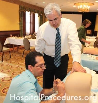
HPC offers a course on lumbar punctures that features hands-on training with simulators and safety training to troubleshoot rare scenarios.
Who Is It For?
The course is aimed at medical professionals who are looking to receive targeted training on lumbar punctures and become subject matter experts.
The course teaches proper techniques for obtaining CSF through a lumbar puncture procedure in the lateral decubitus position and the sitting position.
It covers the proper technique for measuring the opening pressure in the lateral decubitus position. Students can also expect to receive instructions on managing complications that may occur during the procedure.
The course also discusses the indications for lumbar puncture in patients receiving antiplatelets, antithrombotics, or anticoagulant medications.
The Lumbar Puncture course by HPC is part of the live Hospitalist and Emergency Procedures CME course which teaches clinicians how to perform 20 essential procedures for working in emergency, ICU, and hospital settings. It includes King tube placement, laryngeal mask airway (LMA), ultrasound-guided peripheral IV access, and more.
Visit the HPC website for additional information.
Stiffler, K. Scott,S. Robinson,A . The use of ultrasound to identify pertinent landmarks for lumbar puncture. Emerg Med (2007); 25(3) 331-4.
Peterson,M. Abele, J. Bedside ultrasound for difficult lumbar puncture. Emerg Med. 2005; 28 (2); 197-200
Nomura, J. Leech, S. Shenbagamurthi,S. Sierzenski,P. O’Connor, R. Bollinger, M. Humphrey, M. Gukhool, J. A randomized controlled trial of ultrasound-assisted lumbar puncture. J Ultrasound Med 2008; 26(10); 1341-8