This is beneficial when timely diagnosis is essential to treat the patient quickly and appropriately and can help improve patient outcomes. The RUSH exam offers a quick and well-defined evaluation of the condition, allowing healthcare professionals to identify and address the issue based on accurate data.
Knowing the protocol for the POCUS RUSH exam is crucial for healthcare professionals working in the emergency department. This is because quickly identifying the cause of the shock and effectively treating it can significantly impact the final outcome for the patient.
Let’s explore all there is to the POCUS RUSH exam!
What is a POCUS RUSH Exam?
POCUS stands for “point-of-care ultrasound” while RUSH refers to “rapid ultrasound in shock and hypotension.”
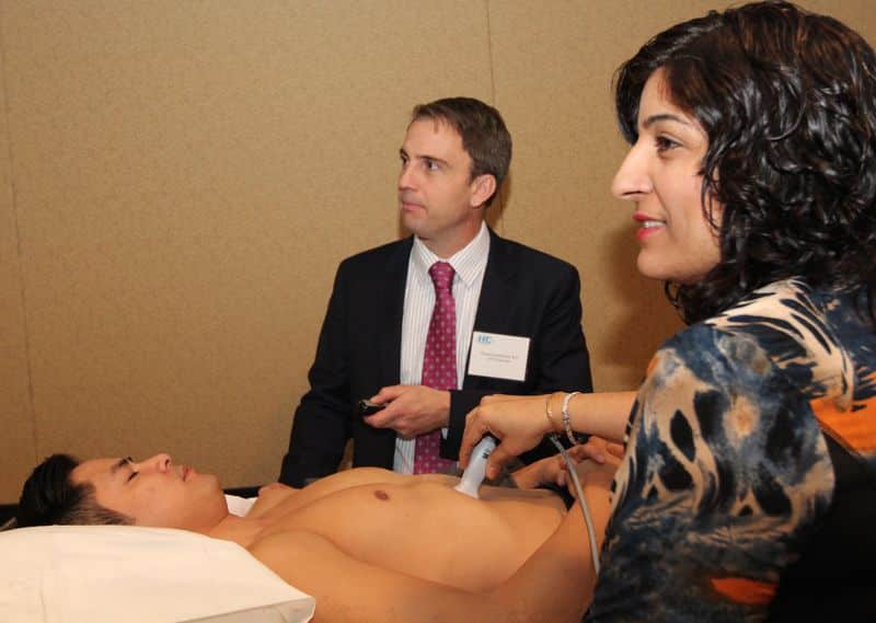
The POCUS RUSH exam is a targeted ultrasound examination used in critical healthcare settings to quickly assess and diagnose the cause of shock or hypotension in a patient. In the emergency department, intensive care unit and hospital wards, this is extremely crucial as professionals have to make immediate decisions. Point-of-care ultrasound can offer real-time information to guide evaluation, treatment and prognosis.
The POCUS RUSH exam involves the use of a point-of-care ultrasound to evaluate the major blood vessels, heart, lungs, abdomen, inferior vena cava (IVC), and other potential sources of hypotension.
Clinicians perform this examination to quickly ascertain the cause of the patient’s symptoms such as hypovolemia, pulmonary embolism, cardiac dysfunction, bleeding, and more.
By identifying the root cause of the symptoms, healthcare professionals can determine the most appropriate course of treatment to address the issue and manage the patient’s condition.
What Is the POCUS RUSH Exam Protocol?
Performed when a patient presents with shock or hypertension, the RUSH protocol is a structured point-of-care examination that is more detailed than the FAST scan. The FAST exam is part of the RUSH exam.
It is conducted by a trained clinician to differentiate between distributive (septic, hypovolemic, hemorrhagic and neurogenic), cardiogenic, and obstructive shock. The protocol involves an assessment of the heart, lungs, and blood vessels.
The heart is assessed for:
- Signs of tamponade
- Signs of strain on the right ventricular
- Pulmonary embolism
- Contractility of the left ventricular
- Pericardial effusion
- Ejection fraction assessment
- Cardiac output assessment
The lungs are evaluated for:
- Pleural effusions
- Tension pneumothorax
- Pulmonary edema
The abdomen is evaluated for:
- Free intraabdominal fluid
- Volume responsiveness as determined by IVC diameter and distensibility
Additionally, the vessels are checked for:
- Dissection of thoracic aortic aneurysm
- Lower limb deep vein thrombosis
- Abdominal aortic aneurysm
- Aortic dissection
After assessing these areas and checking for abnormalities, the clinician may be able to ascertain the cause and type of shock that the patient is experiencing as each has its own features.
Hypovolemic Shock
Hypovolemic shock is characterized by a lack of blood in the blood vessels, usually caused by blood loss.
Blood vessels carry vital nutrients and oxygen to the organs while ensuring that the organs are functioning properly and do not fail as a result of hypoperfusion. In case of hypovolemic shock, they are unable to deliver the required oxygen to the organs, causing them to malfunction ultimately fail.
The main features of hypovolemic shock evaluable with ultrasound include:
- Hypercontractile heart
- Flat Internal Jugular vein and Inferior Vena Cava
- Peritoneal or pleural blood
- Aortic dissection
- Ruptured AAA
Patients experiencing severe dehydration may also experience hypovolemic shock.
Cardiogenic Shock
Cardiogenic shock can be determined by looking for noticeable features such as:
- Dilated heart
- Distended IVC and IJ
- Pleural or peritoneal effusions or ascites
- Echogenic B-lines in the lung caused by a pulmonary edema
- Hypocontractile heart
Cardiogenic shock occurs when the heart is damaged, thus reducing the blood flow in the body. It can also be due to an irregular or slow heart rhythm.
Obstructive Shock
Obstructive shock, as the name suggests, occurs when the flow of blood is obstructed in any way.
Features visible in an ultrasound that indicate an obstructive shock include:
- RV strain
- DVT
- Pneumothorax
- Distended IJ vein and IVC
- Pericardial effusion
- Hypercontractile heart
The blood flow may be interrupted due to pulmonary embolism, or other conditions that cause air or fluid to build up in the chest cavity such as a hemothorax or cardiac tamponade.
Distributive Shock
Distributive shock occurs when the blood vessels lose their tone which can make them open, floppy, and unable to supply enough oxygen and blood to the organs. Findings of distributive shock on POCUS include:
- Peritoneal fluid
- Pleural fluid
- Flat or normal IVC and Internal Jugular vein
- Hypercontractile heart during early sepsis
- Hypocontractile heart during the late stages of sepsis
Distributive shock may be caused by a severe allergic reaction, blood poisoning, damage to the central nervous system, brain injuries, or drug toxicities.
Final Thoughts
The POCUS RUSH exam can offer crucial information on what is causing a patient to experience shock, be it cardiogenic, distributive, obstructive, or hypovolemic shock. The clinician can then effectively treat the underlying cause with the information gathered and determine the best course of treatment.
It can also provide valuable information by allowing healthcare professionals to understand how shock can be prevented since it offers insights into the patient’s cardiovascular health.
To provide effective medical care during critical situations, it is, therefore, important to know what the protocol for a POCUS RUSH exam is.
Learn more about the procedure from the RUSH Exam Course offered by Hospital Procedures Consultants. This course will teach you how to efficiently identify the cause of shock through the proper and thorough examination of its results.
Resources
Rice, J. Brewer, J. Speaks, T. Choi, C. Lahsei, P. Romito, B. The POCUS Consult: How Point of Care Ultrasound Helps Guide Medical Decision Making. Int J Gen Med. 2021; 14: 9789–9806.
Seif, D. Perera, P. Mailhot, T. Riley, D. Mandavia, D. Bedside Ultrasound in Resuscitation and the Rapid Ultrasound in Shock Protocol. Crit Care Res Pract. 2012; 2012: 503254.