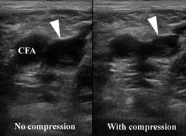
Frequently the combination of a serum D-dimer level and compression ultrasound testing can differentiate between a residual venous clot and an acute recurrent DVT. However, sometimes the results are equivocal and in these situations an MRI direct thrombus imaging can help distinguish a residual clot from a recurrent DVT.
To examine the usefulness of magnetic resonance direct thrombus imaging (MRDTI) in this setting, investigators in the Netherlands performed a prospective, multicenter study involving 39 patients with symptomatic, acute-recurrent DVT. The researchers also studied 42 asymptomatic persons who had previous history of DVT, ultrasonic evidence of chronic residual thrombosis, and a normal d-dimer test. The mean age of patients was 52, 64% were male, and the median time after the previous DVT was 83 months. All individuals received MRDTI that was interpreted by observers blinded to patient status.
MRDTI results were abnormal in 95% of patients with acute recurrent DVT and normal in all 42 asymptomatic patients; the sensitivity and specificity of MRDTI in these patients were 95% (95% confidence interval, 83%–99%) and 100% (95% CI, 92%–100%), respectively.
Reference: